BLOG

IMPETUX releases Deimus T-10i into the market!

IMPETUX releases Deimus T-10i into the market!

The IMPETUX team is glad to announce the launch of the entry-level optical force measurement system: Deimus T-10i.

As a result of our continuous development activity we are proud to present our new T series system for Optical Tweezers

Do you want to multiply the possibilities of your optical trapping setup at a low price?

Do you want to finally achieve those quantitative results you have been looking for quickly and easily?

Deimus T-10i is an entry-level version that can fulfill all your needs.

Impetux’s exclusive, patented technology, leads you where other force methods cannot. Our products represent a change in the force measurement field, making possible and simple the experiments where traditional trap stiffness calibration fails.

Deimus T-10i represents:

  • Novelty: It allows the measurement of optical forces in experiments where the calibration is complicated or impossible
  • Versatility: it is compatible with different microscope models
  • Affordability: excellent price performance ratio

Find out now how Deimus T-10i can lead you further on your research.
More news:

Fig. 1

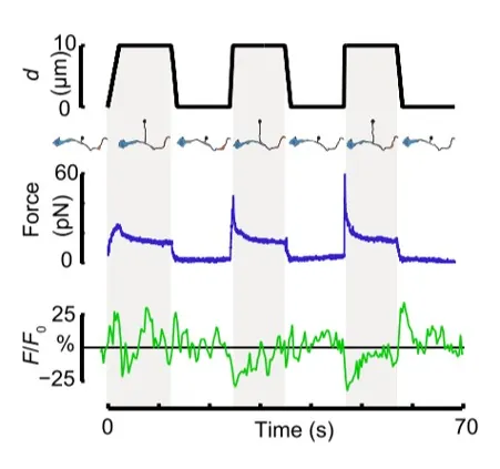
Time dependence of trap position (top), force signal (middle) and Ca2+ fluorescence signal (bottom) recorded during a membrane tether pulling experiment. The force and Ca2+ ion channel activity signals exhibit negative correlation.

Fig. 2

Force and displacement data for two beads stimulated at frequencies of 4 Hz and 0.25 Hz. The progressive reduction of the beads displacement is in concomitancy with an increase of the applied force and loading rate.

Fig. 1

Schematic representation of the optical tweezers experiment. A fibronectin-coated bead is trapped, brought into contact with the cell membrane and stimulated with oscillations at different frequencies.

Fig.2

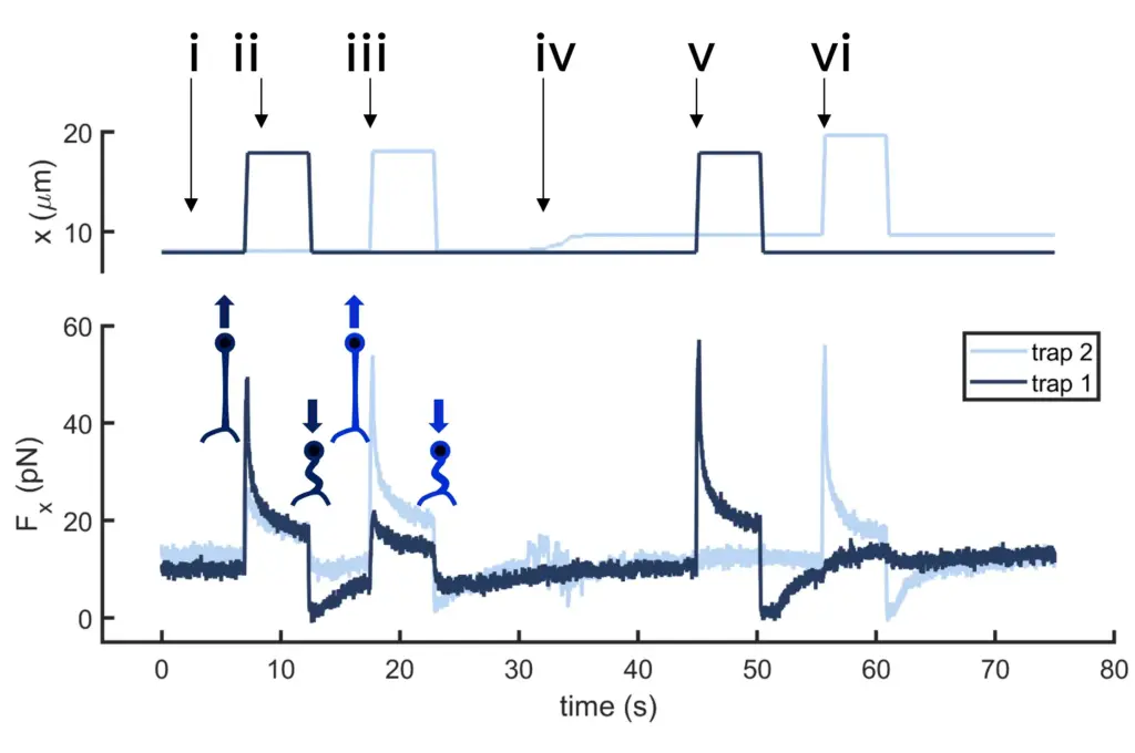
Time dependence of trap 1 and trap 2 position (top) and force (bottom) data along the different steps of the described dual tether pulling experiment.

Fig.1

Confocal video of the described dual tether pulling assay performed on a neuron axon. Courtesy of M. Krieg lab (ICFO).

Fig. 1

Time dependence of trap position (top), force signal (middle) and Ca2+ fluorescence signal (bottom) recorded during a membrane tether pulling experiment. The force and Ca2+ ion channel activity signals exhibit negative correlation.

Video 1

Confocal video showing a DVA neuron Ca2+ ion channel activity in a dynamic optical trapping assay. The pulling rate applied to the membrane tether is progressively increased. Scale bar = 5µm. Acquired at 10Hz.