BLOG

Optical Tweezers: Methods and Protocols

Optical Tweezers: Methods and Protocols

We have the pleasure to announce the publication of the book:

Optical Tweezers: Methods and Protocols

llibreAn indispensable manual edited by Dr Arne Gennerich that includes cutting-edge methods and protocols, provides step-by-step detail essential for reproducible results and contains key notes and implementation advice from the experts.

Among its 21 chapters full of invaluable and great contributions from the most reputable experts around the world, you will find our humble contribution on chapter 3rd: Beyond the Hookean Spring Model: Direct Measurement of Optical Forces Through Light Momentum Changes written by Arnau Farre, Ferran Marsa  and Mario Montes-Usategui.

Enjoy the reading!
More news:

Fig. 1

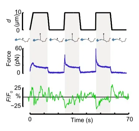
Time dependence of trap position (top), force signal (middle) and Ca2+ fluorescence signal (bottom) recorded during a membrane tether pulling experiment. The force and Ca2+ ion channel activity signals exhibit negative correlation.

Fig. 2

Force and displacement data for two beads stimulated at frequencies of 4 Hz and 0.25 Hz. The progressive reduction of the beads displacement is in concomitancy with an increase of the applied force and loading rate.

Fig. 1

Schematic representation of the optical tweezers experiment. A fibronectin-coated bead is trapped, brought into contact with the cell membrane and stimulated with oscillations at different frequencies.

Fig.2

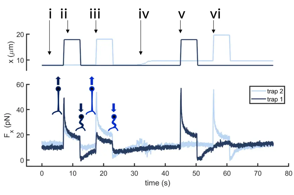
Time dependence of trap 1 and trap 2 position (top) and force (bottom) data along the different steps of the described dual tether pulling experiment.

Fig.1

Confocal video of the described dual tether pulling assay performed on a neuron axon. Courtesy of M. Krieg lab (ICFO).

Fig. 1

Time dependence of trap position (top), force signal (middle) and Ca2+ fluorescence signal (bottom) recorded during a membrane tether pulling experiment. The force and Ca2+ ion channel activity signals exhibit negative correlation.

Video 1

Confocal video showing a DVA neuron Ca2+ ion channel activity in a dynamic optical trapping assay. The pulling rate applied to the membrane tether is progressively increased. Scale bar = 5µm. Acquired at 10Hz.