
BLOG

Lunam T-40i application for force sensing with geometrically anisotropic probes

Lunam T-40i application for force sensing with geometrically anisotropic probes

“Optical Force Sensing With Cylindical Microcontainers”

Recently, the paper: “Optical Force Sensing With Cylindical Microcontainers” has been published in the Particle & Particle System Characterization Journal.

In this work Professor Denz and her team: Mr. Meissner and Dr. Oliver, use our force measurement system commercially available:  Lunam T-40i, to measure forces on cylindrical probes (*) in order to establish whether the established methodology for force sensing based on spherical objects still applies for other geometrically anisotropic probes.

(*) “there is an increasing demand in using force probes in elongated or cylindrical shape: Most biological probes that are of utmost importance to guarantee biocompatibility of in vivo force metrology, are no spherical objects”.

Congratulations for the results team !.

You will find the complete paper here.

You will find more information about our commercially available instruments for Biophysics and Mechanobiology here.
 

More news:

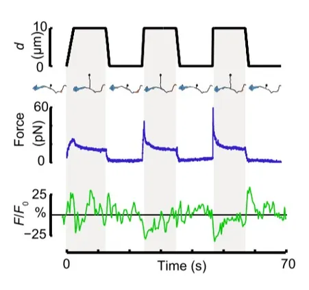
Fig. 1

Time dependence of trap position (top), force signal (middle) and Ca2+ fluorescence signal (bottom) recorded during a membrane tether pulling experiment. The force and Ca2+ ion channel activity signals exhibit negative correlation.

Fig. 2

Force and displacement data for two beads stimulated at frequencies of 4 Hz and 0.25 Hz. The progressive reduction of the beads displacement is in concomitancy with an increase of the applied force and loading rate.

Fig. 1

Schematic representation of the optical tweezers experiment. A fibronectin-coated bead is trapped, brought into contact with the cell membrane and stimulated with oscillations at different frequencies.

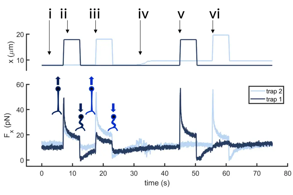
Fig.2

Time dependence of trap 1 and trap 2 position (top) and force (bottom) data along the different steps of the described dual tether pulling experiment.

Fig.1

Confocal video of the described dual tether pulling assay performed on a neuron axon. Courtesy of M. Krieg lab (ICFO).

Fig. 1

Time dependence of trap position (top), force signal (middle) and Ca2+ fluorescence signal (bottom) recorded during a membrane tether pulling experiment. The force and Ca2+ ion channel activity signals exhibit negative correlation.

Video 1

Confocal video showing a DVA neuron Ca2+ ion channel activity in a dynamic optical trapping assay. The pulling rate applied to the membrane tether is progressively increased. Scale bar = 5µm. Acquired at 10Hz.