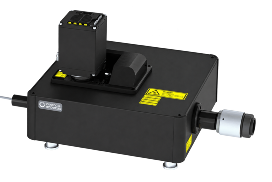
PhaseStreak brings a solid-state approach to line-scanning confocal microscopy, enabling fast, deep imaging with direct control over illumination and optical sectioning.
SOLID-STATE ARCHITECTURE
No moving parts or physical pinholes
230fps · 23.1mm FOV · 300+µm DEEP
Fast, large-field and deep confocal imaging
GENTLE LIVE-CELL IMAGING
Efficient illumination and strong background rejection reduce light exposure
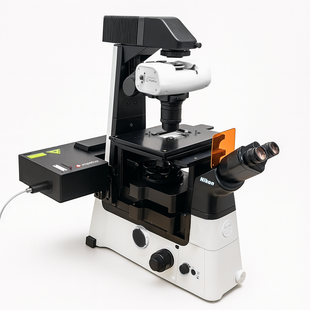
A flexible platform combining programmable optical trapping, direct force measurement and fluorescence imaging to study molecular interactions, cellular mechanics and complex soft matter.
PhaseStreak brings a solid-state approach to line-scanning confocal microscopy, enabling fast, deep imaging with direct control over illumination and optical sectioning.
SOLID-STATE ARCHITECTURE
No moving parts or physical pinholes
230fps · 23.1mm FOV · 300+µm DEEP
Fast, large-field and deep confocal imaging
GENTLE LIVE-CELL IMAGING
Efficient illumination and strong background rejection reduce light exposure
A flexible platform combining programmable optical trapping, direct force measurement and fluorescence imaging to study molecular interactions, cellular mechanics and complex soft matter.
Explore our expertise in live-cell imaging and quantitative optical trapping.
Designed to follow biology in space and time
PhaseStreak combines fast optical sectioning, efficient illumination and deep-sample imaging to support demanding live-cell and 3D fluorescence workflows.
01
FAST 3D IMAGING
Rapidly acquire volumetric fluorescence datasets.
02
DEEP-SAMPLE IMAGING
Maintain optical sectioning and background rejection across thick samples.
03
DESIGNED FOR LONG-TERM IMAGING
Optimize sectioning and signal collection while illuminating only the region of interest.
04
FOUR-COLOR IMAGING
Sequential acquisition at 405, 473, 561 and 639 nm.
Probe the forces shaping living and soft-matter systems
Use optical trapping to investigate molecular interactions, cellular mechanics, biomolecular condensates and the physical properties of complex soft matter.
01
MECHANOBIOLOGY
Investigate force generation, viscoelasticity and mechanotransduction in cells, tissues and living organisms.
02
MOLECULAR BIOPHYSICS
Quantify molecular interactions and the dynamics of individual biomolecules, molecular motors and cytoskeletal filaments.
03
BIOMOLECULAR CONDENSATES
Investigate the mechanics, viscoelasticity and fusion dynamics of biomolecular condensates formed through liquid-liquid phase separation.
04
SOFT MATTER PHYSICS
Characterize colloidal interactions and the rheological properties of complex and active materials
PhaseStreak combines fast optical sectioning, efficient illumination and deep-sample imaging to support demanding live-cell and 3D fluorescence workflows.
01
FAST 3D IMAGING
Rapidly acquire volumetric fluorescence datasets.
02
DEEP-SAMPLE IMAGING
Maintain optical sectioning and background rejection across thick samples.
03
DESIGNED FOR LONG-TERM IMAGING
Optimize sectioning and signal collection while illuminating only the region of interest.
04
FOUR-COLOR IMAGING
Sequential acquisition at 405, 473, 561 and 639 nm.
Probe the forces shaping living and soft-matter systems
Use optical trapping to investigate molecular interactions, cellular mechanics, biomolecular condensates and the physical properties of complex soft matter.
01
MECHANO -BIOLOGY
Investigate force generation, viscoelasticity and mechanotransduction in cells, tissues and living organisms.
02
MOLECULAR BIOPHYSICS
Quantify molecular interactions and the dynamics of individual biomolecules, molecular motors and cytoskeletal filaments.
03
BIOMOLECULAR CONDENSATES
Investigate the mechanics, viscoelasticity and fusion dynamics of biomolecular condensates formed through liquid-liquid phase separation.
04
SOFT MATTER PHYSICS
Characterize colloidal interactions and the rheological properties of complex and active materials
“The IMPETUX team has been an excellent partner throughout system specification, installation, and training, and continues to provide dedicated support for a wide range of projects and assay-protocol development.”
“Thanks to IMPETUX, we have been able to make some very exciting discoveries, resulting in a series of key publications in Nature Physics, Nature Materials, and other leading journals. But what really makes the difference is the support we receive. You really feel that they genuinely care about the science and the research we are doing”
“IMPETUX has been an outstanding collaborator in developing and applying optical tweezers to challenging problems in biological physics. Their highly capable, flexible instrumentation, together with their exceptional technical support and collaborative approach, has enabled us to develop new experimental methods for probing the mechanics of living cells, biomolecular condensates, and other active matter systems."
“Impetux SENSOCELL is the perfect instrument for our research. Having used multiple optical traps in my career, I am impressed by its abilities and most importantly ease of use. This is critical to make optical trapping accessible to the whole lab. We’ve also had a great time interacting with the folks at Impetux who have been altogether very helpful and supportive”
DISTINCTIVE PHOTONIC TECHNOLOGIES
Solid-state confocal imaging and calibration-free optical tweezers.
BORN FROM RESEARCH
Engineered in Barcelona at the intersection of photonics, physics and biology.
VERSATILITY BY DESIGN
Multiple samples, workflows and research questions. One instrument.
SCIENTISTS SUPPORTING SCIENTISTS
Work directly with our team as your experiments evolve.
LATEST FROM IMPETUX
News & Publications
LAUNCH
Sept 2025
PhaseStreak Confocal — now available
Our second instrument platform combines quantitative phase imaging with high-speed confocal fluorescence.
Read more →
PUBLICATION
July 2025
Novel actin polymerization force regime discovered with SENSOCELL
Collaborators at Institut Curie publish new findings in Nature Cell Biology.
Read more →
EVENT
May 2025
Impetux at EBSA 2025 — Booth 42, Barcelona
Live demos and a first look at PhaseStreak Confocal. Book a meeting with our scientists.
Read more →
GET IN TOUCH
Talk to one of our scientists
Tell us about your research, samples or experimental requirements. Our team will help you explore the right optical trapping or confocal imaging solution.
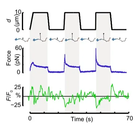
Time dependence of trap position (top), force signal (middle) and Ca2+ fluorescence signal (bottom) recorded during a membrane tether pulling experiment. The force and Ca2+ ion channel activity signals exhibit negative correlation.
Fig. 2
Force and displacement data for two beads stimulated at frequencies of 4 Hz and 0.25 Hz. The progressive reduction of the beads displacement is in concomitancy with an increase of the applied force and loading rate.
Fig. 1
Schematic representation of the optical tweezers experiment. A fibronectin-coated bead is trapped, brought into contact with the cell membrane and stimulated with oscillations at different frequencies.
Fig.2
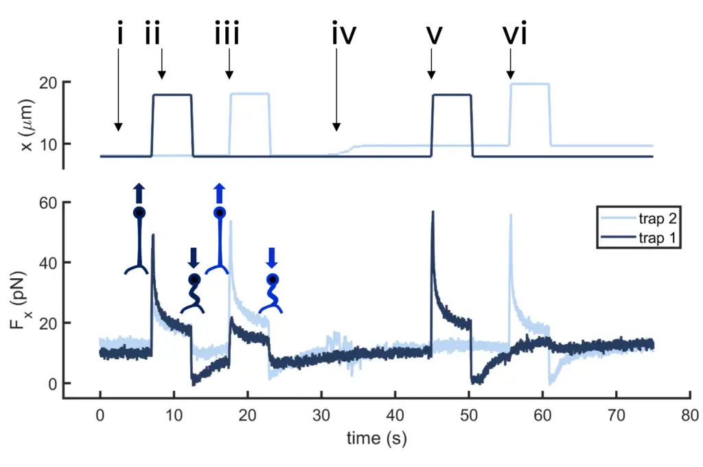
Time dependence of trap 1 and trap 2 position (top) and force (bottom) data along the different steps of the described dual tether pulling experiment.
Fig.1
Confocal video of the described dual tether pulling assay performed on a neuron axon. Courtesy of M. Krieg lab (ICFO).
Fig. 1
Time dependence of trap position (top), force signal (middle) and Ca2+ fluorescence signal (bottom) recorded during a membrane tether pulling experiment. The force and Ca2+ ion channel activity signals exhibit negative correlation.
Video 1
Confocal video showing a DVA neuron Ca2+ ion channel activity in a dynamic optical trapping assay. The pulling rate applied to the membrane tether is progressively increased. Scale bar = 5µm. Acquired at 10Hz.